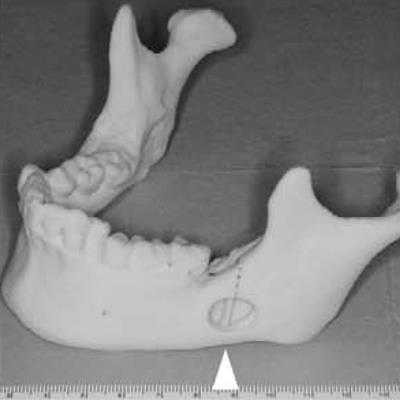
Radiography, CT, and 3D modeling helped clinicians remove burs that had broken off from high-speed handpieces during third-molar extractions and migrated to the mandibular body of one patient and the floor of the mouth of another, according to a recently published report of two cases.
Read more on DrBicuspid.com
 Radiography, CT, and 3D modeling helped clinicians remove burs that had broken off from high-speed handpieces during third-molar extractions and migrated to the mandibular body of one patient and the floor of the mouth of another, according to a recently published report of two cases.
No comments:
Post a Comment